
Biological Energy Transduction
Life depends on constant energy transduction mechanisms. In all free-living organisms, from prokaryotes to eukaryotes, the obtained energy whether from light, inorganic or organic compounds is transduced into a transmembrane difference of electrochemical potential across the prokaryotic cytoplasmatic or mitochondrial membranes. For example, non-photosynthetic organisms obtain energy by the degradation of organic food components, such as proteins, carbohydrates, and lipids, whose metabolisms provide substrates to the respiratory chain. Here chemical reactions are coupled to the translocation of ions across the membrane and the energy thus released by the favorable chemical reaction is transduced to the form of a transmembrane difference of electrochemical potential. This transmembrane potential is vital for solute/nutrient cell transport, synthesis of ATP and motility.
The research in our group focuses on the elucidation of the mechanisms of energy transduction in biological systems. We have been exploring different energy transducing respiratory enzymes, namely their structural features and catalytic characteristics enabling to dissect molecular mechanisms of electron transfer, ion translocation and their coupling. We have been also scrutinizing non-energy transducing respiratory enzymes since these are mostly present in pathogens and may inspire novel drug targets. We perform a multidisciplinary approach using a wide range of molecular biology, biochemical and biophysical methodologies. Our studies involve isolated proteins (wild type and recombinants) as well as reconstituted proteins or membrane vesicles and cell fitness and metabolomic analyses, resulting, in this way, in a synergy of molecular and cellular approaches.
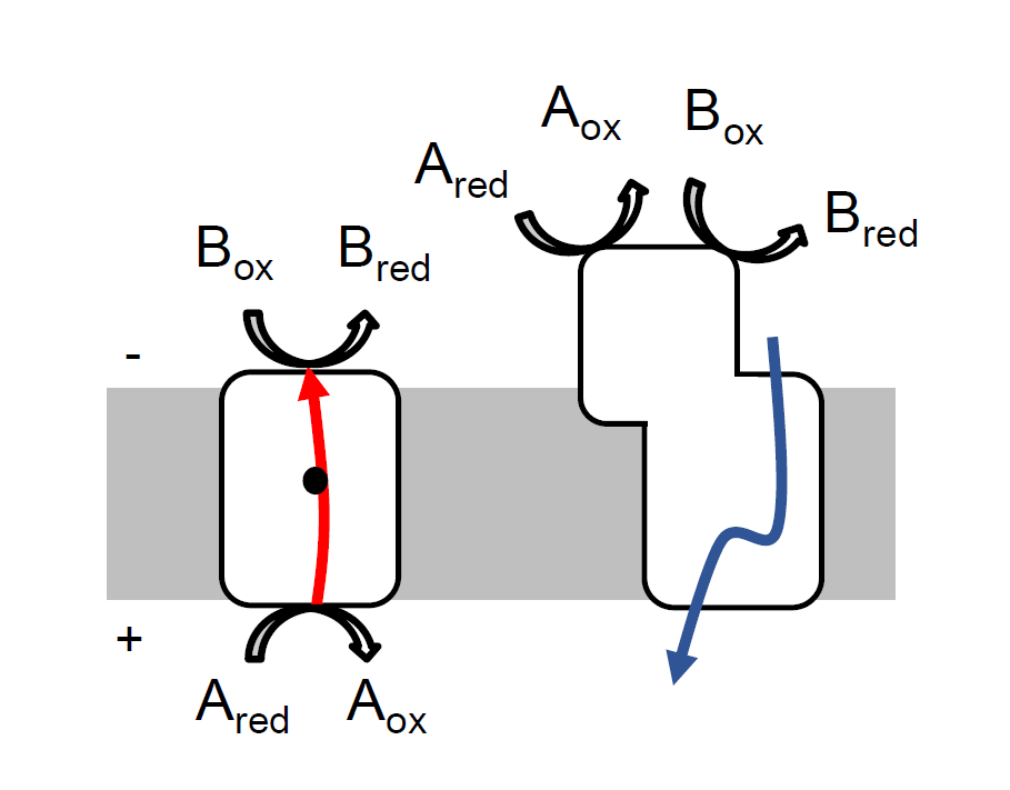
Mechanisms for the establishment of the transmembrane difference of the electrochemical potential ( ). Schematic representation of a cell membrane containing energy transducing membrane protein complexes. All charge-translocating proteins follow two molecular coupling mechanisms: direct- or indirect-coupling (left and right, respectively). In a direct-coupling mechanism the translocated charges are involved in the catalytic reaction and the energy conservation occurs through the movement of these charges, electrons (solid red arrows) and protons (solid blue arrows) across the membrane (grey boxes), against their electrochemical gradient . For electron transfer to be possible, due to the membrane width, the presence of prosthetic groups inside membrane proteins is required (black dot). In an indirect-coupling mechanism, the translocated charges are not involved in the catalytic reaction, which takes place at the periphery of the membrane facing the negative (N, -) or positive -side (P, +) (for clarity only one of these possibilities is schematized in the figure). The Gibbs energy change of the catalytic reaction powers the movement of ions (H+ or Na+) through the membrane from the N- to the P-side of the membrane (d, e, f, g, h).